

Today, we’re taking a deep dive into the fascinating world of X-ray technology. Whether you’re a chiropractor looking to learn more about medical tools, a podiatrist looking to upgrade your imaging equipment, or just someone who wants to learn more about medical technology, we’ve got you covered.
We will explain how X-ray machines work, how images are formed, and how they assist medical professionals in diagnosis and treatment. Our goal is to equip you with the knowledge to make informed decisions in your practice. Get started now!
How Does an X-Ray Machine Operate?
At the heart of every X-ray machine is an X-ray tube, which is similar to a normal light bulb, but more powerful. When electricity is applied, the current heats the filament in the X-ray tube, releasing electrons. These electrons are then accelerated toward a metal target (usually made of tungsten), producing X-rays.
It's a high-speed collision at the atomic level! The X-rays then travel through the body and reach a detector on the other side. Different tissues absorb them at different rates - more in bone, less in soft tissue - creating the image we see. Understanding how these things work is key to using X-ray technology effectively.
How Does an X-Ray Machine Produce an Image?
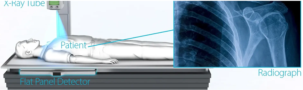
Step 1: The X-ray machine starts the scanning process by generating X-rays. When an electric current heats the filament in the X-ray tube, it emits electrons, which collide with the metal target, producing X-rays.
Step 2: The patient is carefully placed between the X-ray machine and the detector. X-rays pass through the patient's body and reach the detector.
Step 3: Different tissues in the body absorb different amounts of X-rays. Dense structures, such as bones, absorb more X-rays and appear white on the image.
Step 4: Soft tissues, such as muscles and organs, absorb less X-rays and appear as varying shades of gray on the image.
Step 5: Areas containing air, such as the lungs, absorb the least amount of X-rays and therefore appear black on the image.
Step 6: The final image is the result of all these different absorption levels, providing a detailed view of the body's internal structures. This image will become an important tool for diagnosis and treatment.
How Do X-Ray Machines Help Doctors?
X-ray machines are essential aids in helping doctors diagnose, treat and monitor health conditions. They are like eyes that peer into the body, illuminating what lies beneath the surface. Whether it’s an orthopedic surgeon identifying a broken bone or an emergency department quickly diagnosing a potential health crisis, X-rays play a vital role.
More than just a diagnostic tool, they can guide complex procedures such as stent placement or biopsy, providing doctors with real-time images. In addition, the role of X-rays extends to monitoring treatment progress, helping to track how well a fracture heals or how a tumor responds to treatment. Essentially, X-ray machines provide doctors with critical visual data so they can make informed decisions about patient care.
Post time: Jul-14-2025